
LUNG DISEASE
Mediastinoscopy
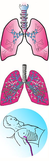
The trachea or wind pipe has lymph nodes running alongside it. These lymph nodes drain the lymph tissue from the lung. Occasionally, lung disease will involve these lymph nodes. In order to determine a diagnosis to aid your doctor in your treatment, we will biopsy one of these nodes. This involves a small incision at the base of your neck and passing a scope to this area to obtain a biopsy.
Lung Biopsy
There are other occasions where we will need to biopsy the lung directly. This can often be performed through the video endoscope. Using general anesthesia, we insert the scope through the chest and inspect the entire chest cavity. The appropriate portion of the lung is selected and a wedge of this tissue can be removed. In addition, fluid and lung cysts can be removed through the same minimally invasive small incision.
Lung Resection
When there is a mass in the lung, we may resect a portion of one lung or occasionally all of the lung. The right lung is divided into three segments, the left into two. Usually, we will remove one of these segments. Before removing any lung, we first inspect to see if the disease process involves the lymph node tissue or the chest wall itself. In some circumstances, we may not remove the lung lesion if the disease has spread elsewhere. After removal of a portion of the lung, the remaining lung will expand to fill the empty space. There will also be small areas of the lung that will leak some air for a few days and then usually stop. As these two processes occur, you will have chest tubes which drain fluid and air and help the lung to re-expand.